Diagnostic Tests

Blood tests
Blood tests cannot be used directly to find or diagnose pancreatic cancer, but they can provide more information about how the pancreas and other parts of your body are functioning.
Some types of pancreatic cancer cells release certain proteins or ‘markers’ into the blood which can be detected in a blood test. After the diagnosis of pancreatic cancer, these markers may help your doctor to determine if your cancer is responding to treatment.
Other medical conditions can raise the levels of these substances and some people with pancreatic cancer will have normal levels of these markers, which is why they cannot be used to diagnose pancreatic cancer and may not always be a useful test.
Markers for pancreatic exocrine tumours (such as pancreatic adenocarcinoma, PDAC) include:
- Carbohydrate associated antigen (CA19-9)
- Carcinoembryonic antigen (CEA)
- CA 125 (cancer antigen)
Markers for pancreatic neuroendocrine tumours (PNETs) include:
Chromogranin A (CgA) is a tumour marker and specific hormones made by the pancreatic neuroendocrine tumours (PNETs), including pancreatic polypeptide (PP) can also be detected through blood tests and used to help diagnose PNETs. Some medications such as protein pump inhibitors (eg. pantoprazole, esomeprazole), can falsely elevate CgA). Patient may be advised by their doctor to withhold these medications prior to performing this test.
Imaging
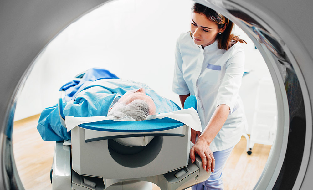
Imaging tests or scans provide visual information about the pancreas and surrounding areas and are essential for diagnosing pancreatic cancer. Different types of scans provide different information, so the type and range of scans is personalised to each individual.
Some of these scans are not available at every hospital and are not always covered by Medicare or private health insurance so it is important to discuss access and healthcare costs with your doctor.
Computer tomography (CT) scan - use x-rays to take several pictures of the inside of the body, which are then compiled into one detailed cross-sectional picture. CTs are often used to determine if the cancer has spread to other organs. With a special contrast injected, CT is also very important to see how the cancer in the pancreas is growing around vital blood vessels and nerves and if confined only to the pancreas, for the surgeon to assess if it can be safely and completely surgically removed.
Ultrasound - uses soundwaves to create a picture of the inside of the body. This may be the first test performed and will particularly help assess whether the bile duct draining from the liver has become blocked potentially by a cancer in the pancreas and whether there has been any spread of the cancer to the liver. This test is very useful in investigating other non-cancerous causes for the symptoms being investigated.
Magnetic resonance imaging (MRI) - uses radio waves and magnets to build a detailed 3D picture of the organs and structures inside the body by measuring their energy. MRI provides very detailed and precise information on the anatomy of the pancreas, surrounding organs and soft tissues and the liver, including if there are any cancer secondaries there.
Magnetic resonance cholangiopancreatography (MRCP) - MRCP scans are a type of MRI that can show detailed images of the pancreatic and bile ducts to find tumours, pancreatic cysts or blockages in the ducts. MRCP is very useful in cases where it is difficult to determine exactly what is happening in the body and more subtle cancers or problems arising from the cancer.
Positron emission tomography (PET) – scans create images based on the chemical reactions happening in cells. Cancer cells will process chemicals, including sugars, differently to normal cells so will show up on the scan. A PET scan may be able to find secondaries or spread of the cancer where other scans including CT have not been able to. It may also be used for research purposes to monitor how the cancer is responding to therapy.
Endoscopic ultrasound (EUS) – uses an ultrasound probe on the end of an endoscope (long thin tube) to take pictures of the pancreas, bile duct and digestive tract. The tube is passed through the patient’s mouth down into the stomach and small bowel to take the images from the inside of the body. This is a common way to biopsy the cancer in the pancreas, as otherwise it is hard to biopsy as sits at the back of the abdomen.
Endoscopic retrograde cholangiopancreatography (ERCP) – uses x-rays to take pictures of the bile duct and, or the pancreatic duct. An endoscope is used to deliver a small amount of dye so that any blockages or narrowing caused by the cancer can be seen on the x-ray. During this procedure, brushings can be taken which may find cancer cells to help diagnosis, infection may be isolated if occurring and if needed a stent can be placed in the bile duct if it is being blocked by the cancer and not able to drain bile from the liver.
Laparoscopy - uses a camera inserted through a small cut in the abdomen to look at the pancreas and surrounding organs. Although different scans provide a lot of information, the abdomen has many organs. Directly visualising these organs including the slippery membrane they are housed in, the peritoneum, is important to assess for any further spread of the cancer around these sites or in the fluid in the abdomen by collecting some washings.
Biopsy
While imaging can help to identify tumours and their location in the pancreas and surrounding organs, collecting a sample of tissue or cells is the only way to confirm if the mass or tumour is cancerous or benign. Collecting the tissue sample is called a biopsy. The tissue sample will then be examined by a pathologist who helps determine the type of pancreatic cancer, including the grade of the cancer, which can indicate how quickly the tumour may grow. Different types of pancreatic cancer are treated differently and also may behave very differently with varying expected prognoses.
A biopsy may be taken during surgery or procedures such as an endoscopic ultrasound (EUS), endoscopic retrograde cholangiopancreatography (ERCP), laparoscopy, or through the skin by a radiologist. Appropriate sedation and numbing of the area being biopsied is always undertaken so it is not unpleasant.
Always consult your doctor or health professional about any health-related matters. PanKind does not provide medical or personal advice and is intended for general informational purposes only. Read our full Terms of Use.
Thank you to the clinicians, researchers, patients, and carers who have helped us create and review our website information and support resources, we could not have done it without you.


